Journal of Northeastern University(Natural Science) ›› 2024, Vol. 45 ›› Issue (7): 921-927.DOI: 10.12068/j.issn.1005-3026.2024.07.002
Previous Articles Next Articles
Ji-hong LIU1( ), Lü-heng ZHANG1, Hai-xu YANG2
Received:2023-03-05
Online:2024-07-15
Published:2024-10-29
Contact:
Ji-hong LIU
About author:LIU Ji-hong,E-mail:liujihong@ise.neu.edu.cnCLC Number:
Ji-hong LIU, Lü-heng ZHANG, Hai-xu YANG. A Saturation Artifact Inpainting Algorithm for Cell Fluorescence Microscopic Images[J]. Journal of Northeastern University(Natural Science), 2024, 45(7): 921-927.
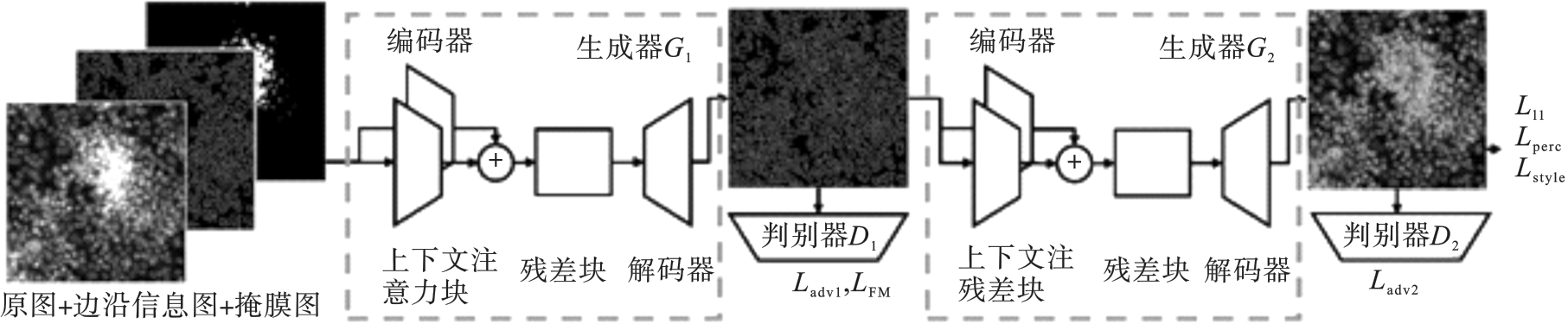
Fig.1 Structure diagram of two stage inpainting model
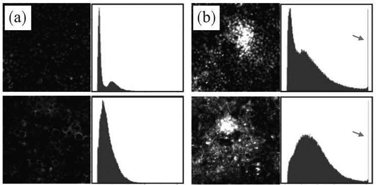
Fig.2 Fluorescence microscopic images of cells
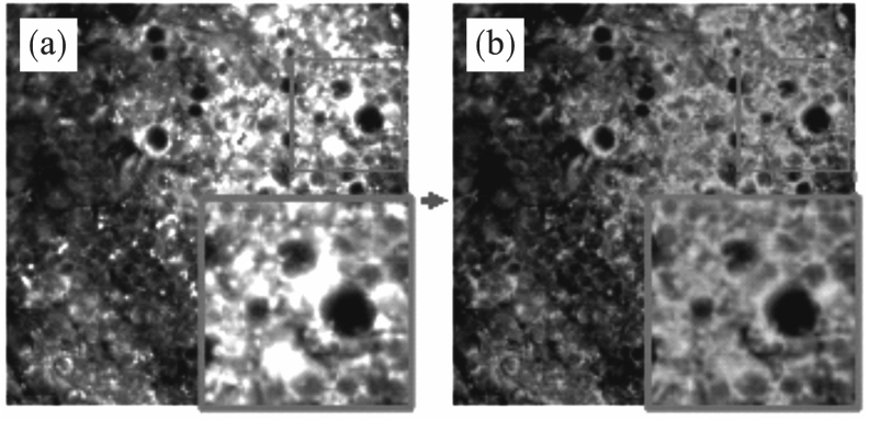
Fig.3 Example of TC-GAN restored results
| 指标 | 数据组 | 均值 |
|---|---|---|
| PSNR | 伪影组 | 9.101 |
| 修复组 | 25.948 | |
| SSIM | 伪影组 | 0.725 |
| 修复组 | 0.854 | |
| FID | 伪影组 | 609.154 |
| 修复组 | 50.345 |
Table 1 Indexes of masked and restored images
| 指标 | 数据组 | 均值 |
|---|---|---|
| PSNR | 伪影组 | 9.101 |
| 修复组 | 25.948 | |
| SSIM | 伪影组 | 0.725 |
| 修复组 | 0.854 | |
| FID | 伪影组 | 609.154 |
| 修复组 | 50.345 |

Fig.4 Qualitative display of TC-GAN restored results
| 测试集 | ACC | SEN | PRE |
|---|---|---|---|
| SET3 | 93.11 | 82.78 | 84.34 |
| SET4 | 92.44 | 81.11 | 83.45 |
Table 2 Indexes of classification on SET3 and SET4
| 测试集 | ACC | SEN | PRE |
|---|---|---|---|
| SET3 | 93.11 | 82.78 | 84.34 |
| SET4 | 92.44 | 81.11 | 83.45 |
| 测试集 | ACC | SEN | PRE |
|---|---|---|---|
| SET5 | 88.22 | 70.56 | 76.75 |
| SET6 | 92.44 | 81.11 | 82.65 |
Table 3 Indexes of classification on SET5 and SET6
| 测试集 | ACC | SEN | PRE |
|---|---|---|---|
| SET5 | 88.22 | 70.56 | 76.75 |
| SET6 | 92.44 | 81.11 | 82.65 |
| 1 | Bougen‑Zhukov N, Loh S Y, Lee H K.et al.Large‑scale image‑based screening and profiling of cellular phenotypes[J].Cytometry:Part A,2017,91(2):115-125. |
| 2 | Caicedo J C, Singh S, Carpenter A E.Applications in image‑based profiling of perturbations[J].Current Opinion in Biotechnology,2016,39:134-142. |
| 3 | Ray K, Ma J, Oram M,et al.Single‑molecule and FRET fluorescence correlation spectroscopy analyses of phage DNA packaging:colocalization of packaged phage T4 DNA ends within the capsid[J].Journal of Molecular Biology,2010,395(5):1102-1113. |
| 4 | Beesley J, Sivakumaran H, Moradi Marjaneh M,et al. eQTL colocalization analyses identify NTN4 as a candidate breast cancer risk gene[J].The American Journal of Human Genetics,2020,107(4):778-787. |
| 5 | Sauvat A, Leduc M, Müller K,et al.ColocalizR:an open‑source application for cell‑based high‑throughput colocalization analysis[J].Computers in Biology and Medicine,2019,107:227-234. |
| 6 | Ren W W, Isler H, Wolf M,et al.Smart toolkit for fluorescence tomography:simulation,reconstruction,and validation[J].IEEE Transactions on Biomedical Engineering,2020,67(1):16-26. |
| 7 | Wang Y, Maslov K, Kim C H,et al.Integrated photoacoustic and fluorescence confocal microscopy[J].IEEE Transactions on Biomedical Engineering,2010,57(10):2576-2578. |
| 8 | Ritter C, Wollmann T, Lee J Y,et al.Data fusion and smoothing for probabilistic tracking of viral structures in fluorescence microscopy images[J].Medical Image Analysis,2021,73:102168. |
| 9 | Caicedo J C, Cooper S, Heigwer F,et al.Data‑analysis strategies for image‑based cell profiling[J].Nature Methods,2017,14(9):849-863. |
| 10 | Moen E, Bannon D, Kudo T,et al.Deep learning for cellular image analysis[J].Nature Methods,2019,16(12):1233-1246. |
| 11 | Bolte S, Cordelières F P.A guided tour into subcellular colocalization analysis in light microscopy[J].Journal of Microscopy,2006,224(3):213-232. |
| 12 | Aladeokin A C, Akiyama T, Kimura A,et al.Network‑guided analysis of hippocampal proteome identifies novel proteins that colocalize with A β in a mice model of early‑stage Alzheimer’s disease[J].Neurobiology of Disease,2019,132:104603. |
| 13 | Zinchuk V, Zinchuk O, Okada T.Quantitative colocalization analysis of multicolor confocal immunofluorescence microscopy images:pushing pixels to explore biological phenomena[J].Acta Histochemica et Cytochemica,2007,40(1):101-111. |
| 14 | Li Y, Xu F, Zhang F,et al.DLBI:deep learning guided Bayesian inference for structure reconstruction of super‐resolution fluorescence microscopy[J].Bioinformatics,2018,34(13):i284-i294. |
| 15 | Ishii S, Lee S, Urakubo H,et al.Generative and discriminative model‑based approaches to microscopic image restoration and segmentation[J].Microscopy,2020,69(2):79-91. |
| 16 | Goodfellow I J, Pouget‑Abadie J, Mirza M,et al.Generative adversarial networks[J].Advances in Neural Information Processing Systems,2014,3:2672-2680. |
| 17 | Tran N T, Tran V H, Nguyen N B,et al.On data augmentation for GAN training[J].IEEE Transactions on Image Processing,2021,30:1882-1897. |
| 18 | Ren Y R, Li G, Liu S,et al.Deep spatial transformation for pose‑guided person image generation and animation[J].IEEE Transactions on Image Processing,2020,29:8622-8635. |
| 19 | Kaneko T, Kameoka H, Hojo N,et al.Generative adversarial network‑based postfilter for statistical parametric speech synthesis[C]//2017 IEEE International Conference on Acoustics,Speech and Signal Processing (ICASSP).New Orleans,2017:4910-4914. |
| 20 | Quan W Z, Zhang R S, Zhang Y,et al.Image inpainting with local and global refinement[J].IEEE Transactions on Image Processing,2022,31:2405-2420. |
| 21 | Chen X J, Zhang C, Zhao J,et al.Weakly supervised neuron reconstruction from optical microscopy images with morphological priors[J].IEEE Transactions on Medical Imaging,2021,40(11):3205-3216. |
| 22 | Belthangady C, Royer L A.Applications,promises,and pitfalls of deep learning for fluorescence image reconstruction[J].Nature Methods,2019,16(12):1215-1225. |
| 23 | Boutros M, Heigwer F, Laufer C.Microscopy‑based high‑content screening[J].Cell,2015,163(6):1314-1325. |
| 24 | Nazeri K, Ng E, Joseph T,et al.EdgeConnect:generative image inpainting with adversarial edge learning[EB/OL]. [2019-01-11].. |
| 25 | Canny J.A computational approach to edge detection[J].IEEE Transactions on Pattern Analysis and Machine Intelligence,1986,8(6):679-698. |
| 26 | Yu J H, Lin Z, Yang J M,et al.Generative image inpainting with contextual attention[C]//2018 IEEE/CVF Conference on Computer Vision and Pattern Recognition. Salt Lake City,2018:5505-5514. |
| 27 | Isola P, Zhu J Y, Zhou T H,et al.Image‑to‑image translation with conditional adversarial networks[C]//2017 IEEE Conference on Computer Vision and Pattern Recognition.Honolulu,2017:5967-5976. |
| 28 | Johnson J, Alahi A, Li F F.Perceptual losses for real‑time style transfer and super‑resolution[C]//European Conference on Computer Vision.Amsterdam,2016:694-711. |
| 29 | Gatys L A, Ecker A S, Bethge M.Image style transfer using convolutional neural networks[C]//2016 IEEE Conference on Computer Vision and Pattern Recognition.Las Vegas,2016:2414-2423. |
| 30 | Russakovsky O, Deng J, Su H,et al.ImageNet large scale visual recognition challenge[J].International Journal of Computer Vision,2015,115(3):211-252. |
| 31 | Bray M A, Singh S, Han H,et al.Cell Painting,a high‑content image‑based assay for morphological profiling using multiplexed fluorescent dyes[J].Nature Protocols,2016,11(9):1757-1774. |
| 32 | Kingma D P, Ba J.Adam:a method for stochastic optimization[EB/OL].[2017-01-30].. |
| 33 | Elharrouss O, Almaadeed N, Al‑Maadeed S,et al.Image inpainting:a review[J].Neural Processing Letters,2020,51:2007-2028. |
| 34 | Heusel M, Ramsauer H, Unterthiner T,et al.GANs trained by a two time‑scale update rule converge to a local Nash equilibrium[EB/OL].[2018-01-12].. |
| 35 | He K M, Zhang X Y, Ren S Q,et al.Deep residual learning for image recognition[C]//2016 IEEE Conference on Computer Vision and Pattern Recognition.Las Vegas,2016:770-778. |
| [1] | Hong-li LI, Hao-yu LIU, Rong-hua ZHANG, Yi CHENG. Emotional Classification Based on Multidimensional Feature Matrix and Improved Dense Connection Network [J]. Journal of Northeastern University(Natural Science), 2024, 45(7): 928-935. |
| [2] | Dong-hong HAN, Yan-ru KONG, Yi-meng ZHAN, Yuan LIU. Research on Emotion Recognition Method of Music Multimodal Data [J]. Journal of Northeastern University(Natural Science), 2024, 45(6): 776-785. |
| [3] | Wei-qi ZHANG, Hui-ming WANG. Interpretable Deep Learning Prediction Model for Compressive Strength of Concrete [J]. Journal of Northeastern University(Natural Science), 2024, 45(5): 738-744. |
| [4] | WEI Jian-yi, WU Jing-jing. Resource Allocation Algorithm in Industrial Internet of Things Based on Edge Computing [J]. Journal of Northeastern University(Natural Science), 2023, 44(8): 1072-1078. |
| [5] | LI Hai-yan, XIONG Li-chang, GUO Lei, LI Hai-jiang. Two-Stage Inpainting Algorithm Based on U-net Edge Generation and Hypergraphs Convolution [J]. Journal of Northeastern University(Natural Science), 2023, 44(3): 331-339. |
| [6] | YANG Xuan, HE Zhan-qi. Improved Two-layer BiLSTM Electrocardiosignal Segmentation Method [J]. Journal of Northeastern University(Natural Science), 2023, 44(12): 1705-1711. |
| [7] | JI Ce, WANG Xin, GENG Rong, LIANG Min-jun. LSTM-Based Channel Estimation Method in Time-Varying Channels [J]. Journal of Northeastern University(Natural Science), 2023, 44(11): 1521-1528. |
| [8] | CHEN Cheng, SHI Pei-xin, WANG Zhan-sheng, JIA Peng-jiao. Shield Load Prediction Method Based on Deep Learning with Multiattention Mechanism [J]. Journal of Northeastern University(Natural Science), 2023, 44(11): 1631-1638. |
| [9] | LI Juan-li, WEI Dai-liang, LI Bo, WEN Xiao. Improved SSD Rapid Separation Model of Coal Gangue Based on Deep Learning and Light-Weighting [J]. Journal of Northeastern University(Natural Science), 2023, 44(10): 1474-1480. |
| [10] | ZHAO Yong, JIAO Shi-hui, ZHAO Qian-bai. Hybrid Recognition Model of Microseismic Signals for Mining Based on Mel Spectrum and LSTM-DCNN [J]. Journal of Northeastern University(Natural Science), 2023, 44(10): 1481-1489. |
| [11] | ZHANG Xue-feng, WANG Zhao-yi. Automatic Lane Change Decision Model Based on Dueling Double Deep Q-network [J]. Journal of Northeastern University(Natural Science), 2023, 44(10): 1369-1376. |
| [12] | WANG Ying, WANG Ze-hao, LI Hong, HUANG Wen-jun. Named Entity Recognition in Threat Intelligence Domain Based on Deep Learning [J]. Journal of Northeastern University(Natural Science), 2023, 44(1): 33-39. |
| [13] | GU De-ying, LUO Yu-lun, LI Wen-chao. Traffic Target Detection in Complex Scenes Based on Improved YOLOv5 Algorithm [J]. Journal of Northeastern University(Natural Science), 2022, 43(8): 1073-1079. |
| [14] | DAI Yin, LIU Wei-bin, DONG Xin-yang, SONG Yu-meng. U-Net CSF Cells Segmentation Based on Attention Mechanism [J]. Journal of Northeastern University(Natural Science), 2022, 43(7): 944-950. |
| [15] | LI Hong-ru, REN Zi-yang, HUANG You-he, YU Xia. Recognition Method of Arrhythmia Based on Variable Weight Singular Spectrum Analysis [J]. Journal of Northeastern University(Natural Science), 2022, 43(3): 305-312. |
| Viewed | ||||||
|
Full text |
|
|||||
|
Abstract |
|
|||||